Wednesday 30 January 2013

Shoulder Arthritis


Dr Senthil Velan MBBS,FRCS (Tr &Ortho), FEBOT, Fellow European Board Orthopaedics and Trauma, D.Ortho, Dip SICOT.

Consultant Orthopaedic Surgeon
Apollo Hospitals, Chennai, India

For appointments contact 09566222533/ velansenthil78@yahoo.com


Shoulder arthritis

What is it?

Arthritis is wear and tear of the joint. Osteoarthritis is a slow process that develops over a number of years. With this type of arthritis the joint cartilage gradually becomes thin and roughened. The bone underneath thickens and the bone at the edge grows outwards causing bony spurs (osteophytes).  The joint may swell as the body makes extra fluid to lubricate the joint. The joint may get deformed and there will be gradual loss of movement.

What is its cause?    

The most common cause of osteoarthritis in the body is wear and tear of the joint due
to ageing. This is called ‘primary osteoarthritis’. Fractures or instability of the shoulder can lead to arthritis later in life. This is termed as ‘secondary arthritis’.

In the shoulder a specific of arthritis known as Rotator cuff arthropathy can develop. It occurs in patients with large, long-standing cuff tears which subsequently develop in to arthritis between the humeral head and acromion. The shoulder is a ball (humeral head) and socket (glenoid) joint. It is raised, lowered or rotated by a group of four muscles (supraspinatus, infraspinatus, subscapularis and teres minor), which are called the ‘rotator cuff muscles’. They also help to stabilize the shoulder joint i.e. they try to keep the ball at the centre of the socket. In the presence of a cuff tear, the ball of the shoulder (Humeral head) will become unstable and move upwards towards the acromion(Shown in Figure below). With time this may cause arthritis between the humeral head and the acromion (tip of the shoulder blade).

X ray showing Shoulder Osteoarthritis


What are the symptoms and how is the condition diagnosed?

The symptoms are of gradually increasing pain and stiffness. With time the symptoms
increase and cause significant functional disability. The patient may complain of
grinding or crunching within the shoulder. In patients with rheumatoid arthritis the shoulder can be damaged at any age. Patients with rheumatoid disease may
have involvement of the other joints in the limb. 

X ray showing Rotator cuff arthropathy

             
What investigations will be needed?

An x-ray of the shoulder is needed. It will confirm the extent and type of arthritis.
A CT scan may be advised to assess loss of bone due to arthritis, to decide
on the right type of replacement. A MR scan may be advised to assess the integrity of
the rotator cuff. 


What is the treatment?

1. Initial management consists of painkillers, anti-inflammatory medication and
activity modification. It is aimed at easing the pain and regaining motion. 

2. If the above measures fail then surgery is considered. The type of surgery that
will depend on the nature and longevity of the symptoms, type and extent of arthritis, age and functional demands of the patient. 

Arthroscopic (keyhole) surgery: A relatively small operation during which a
Telescope is passed into the shoulder through one small incision (½ cm).
Additional 1-2 small stab incisions are used to pass instruments, to allow for
removal of loose bodies, inflammatory tissue and debride any loose cartilage.
Such arthroscopic debridement will provide temporary  relief. 

Shoulder replacement: The eventual solution to an arthritic shoulder is to replace
it. Different types of shoulder replacements can be used, the decision of which is
based on age, functional demands of the patient, extent of arthritis, any bony loss
and presence of a rotator cuff tear. 

Surface replacement: In this type of shoulder replacement, just the humeral head
is replaced. The advantage of this procedure is that it preserves the maximum
amount of bone and minimal soft tissue dissection is needed. If the replacement
fails in the long-term (all replacements will eventually) revision surgery to change
the prosthesis is easier.

Surface Replacement Procedure


Total shoulder arthroplasty: Both the socket (glenoid) and the ball humeral head
are replaced.


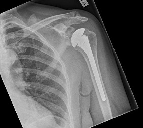
Reverse geometry total shoulder replacement: This procedure in which both the
ball and the socket are replaced and reversed, is practiced for rotator cuff
arthropathy. In the presence of a cuff tear, the deltoid is the only functioning
muscle. Reversing the normal shoulder medializes the centre of rotation of the
joint. This maximises the lever arm of the deltoid muscle and helps achieve a
better range of motion of the shoulder.


Reverse Geometry replacement


What happens if it is not treated?

It is possible that with time pain may lessen, but motion between the humeral head
and glenoid will be lost. However most patients will find that their symptoms will
increase with time and they will need further treatment


What is the success of surgical treatment?

Each of the surgical treatments is successful if they have been advised at an
appropriate stage of the disease. More than 90% patients will achieve a significant
improvement in their pain after these procedures. Success rates for regaining the
movement will vary and you may want to discuss this further with your Shoulder
Specialist. The strength in your arm will take longer to improve, and will be dependent  on the amount of pain and stiffness you had prior to the surgery.


What are the complications of surgical treatment?

1. The surgical scar may remain unattractive. An area of numbness can occur around
the scar but this does not cause any problems. 
2. Infection of the wound is possible but usually can be successfully treated with
antibiotics. Very rarely infection may spread into the joint replacement. 
3. Injury to the nerves (axillary) can occur. This may lead to altered sensations and
shoulder / limb weakness.
4. The shoulder replacement may dislocate.  
5. In the long term, the shoulder joint replacement may fail. In this case, the
replacement will need to be revised. It is expected that nearly 85-90% of shoulder
replacements will still be successful after 10 years.  
6. The replacement may fail to relieve the pain totally. 
7. Shoulder stiffness
8. Fracture of the glenoid or humerus
9. Any surgical intervention has the risk of developing complications / setbacks
which are unpredicted. These complications may have the potential to leave the
patient worse than before surgery. 

Is there anything I can do to improve the outcome?

• Keep the wounds dry and clean until they have healed.
• After the operation the therapist will see you to start movements of the shoulder.
It is important to carry out the prescribed exercises regularly, both during the
physiotherapy sessions and at home. It will help to keep the pain levels down with
analgesics so as to keep your shoulder, elbow and hand fingers moving. 
• It is advised against wearing rings on the operated side for 4 to 6 weeks after
surgery. 

When can I do various activities?

Return to work depends on many factors including the nature of the job and hand
dominance. Following shoulder replacement you will be to return to a desk job within
4-6 weeks of the operation and perform reasonable tasks with the limb by that time. 
Manual work is not generally recommended after a shoulder replacement. 
Driving should be possible within 1 or 2 weeks of keyhole surgery, and within 4 to 6
weeks of shoulder replacements. Before driving, do check that you can manage all
controls and start with short journeys.  

Saturday 26 January 2013

Shoulder Instability (Recurrent Dislocation)


Dr Senthil Velan 
MBBS, MRCS(UK) FRCS (Tr &Ortho), FEBOT(France), Fellow European Board Orthopaedics and Trauma., D Ortho,Dip SICOT(Honk Kong).

Consultant Orthopaedic Surgeon
Apollo Hospitals Ayanambakkam , Chennai, India
For appointments and queries contact us on  Ph 9566222533/ nexusortho@yahoo.com

Shoulder dislocation and instability.

What is it?

The shoulder joint is a ball and socket joint. The round end of the upper arm
bone (humerus) fits into the shallow socket (glenoid) on the shoulder blade
(scapula) much like a golf ball rests on a tee. The anatomy of the shoulder
joint makes it the most mobile joint in the body.

While this gives us the ability to move it in many directions it also makes it an easy joint to dislocate.  A complete dislocation means that the ball (humeral head) has come all the way out of the socket. When the ball comes out of the socket incompletely, it is called a partial dislocation or subluxation. When the shoulder dislocates or subluxes repeatedly it is known as shoulder instability. The shoulder joint can dislocate forward, backward  or downward. The most common type is when the shoulder slips forward (anterior).
                                       
What is its cause?

A ring of cartilage surrounds the shallow socket of the shoulder joint (glenoid).
This ring is called the labrum. The glenoid labrum increases the depth of the
socket making the shoulder joint more stable. A tough, but thin elastic tissue
(capsule) further surrounds the shoulder like a bag. The capsule contains a
series of ligaments or bands, which connect the humerus to the glenoid. 

Labrum deepens the glenoid to provide more stability.
                                                    

The most common cause for shoulder dislocation is a forceful injury like falling
on an outstretched hand (sports injuries, falls) or a hard blow to the shoulder
(a cricket injury or a road traffic accident). This is called a traumatic
dislocation.

 X rays showing dislocation of shoulder joint


The force that causes this injury can cause the labrum to be torn
from the socket making it shallow. This lesion is known as a Bankart’s lesion
and it can lead to further episodes of subluxation or dislocation. 

Sometimes a relatively minor force may dislocate the shoulder for the first
time and the patient may feel a pop as it reduces by itself. This will occur in
people who have normal but lax joints. This is referred to as an atraumatic
dislocation.

A very small group of patients can dislocate their  shoulders without any
trauma. These type of dislocations / subluxations occur because of abnormal
working of the muscles around the shoulder. Such a condition is referred to as
‘positional instability of the shoulder’.
  
What are the symptoms and how is it diagnosed?

When the shoulder dislocates the first time after an injury, the patient will
immediately complain of pain, deformity and weakness in the shoulder. The
patient may develop bruising and complain of numbness or tingling if any of
the nerves around the shoulder have stretched.  
The symptoms of shoulder instability will range from obvious repeated
dislocations, to just pain with certain activities or shoulder positions. Patients
may complain of clicking or popping of the shoulder, the arm suddenly ‘feeling
dead’ or the shoulder been generally weak. 

MRI Scan of shoulder showing labral injury
                                            

    Arthroscopic picture showing Hill Sachs lesion
                                     .

Will further tests or investigations be needed?

After appropriate clinical assessment, an x-ray of  the shoulder is
recommended. When the shoulder is dislocated the first time an x-ray will
reveal any broken bone.   In some situations after the first injury, or if the first injury progresses to  instability, a MR Arthrogram is advised. A dye is injected into the shoulder  and a magnetic field is used to create cross sectional images of the shoulder.
Sometimes a CT scan may be recommended to assess the bony defects. 
Electrical tests may also be necessary if the patient has tingling, numbness or
an altered sensation, to assess for any nerve damage.
                                       
MR arthrogram  image showing  anterior labral  injury (Bankart  lesion)
                             

What is the treatment?

1. After the first dislocation the shoulder will need to be reduced usually in
the Accident & Emergency department. This will be carried out using a
muscle relaxant, sedative or rarely a general anaesthetic. Following
reduction of the shoulder you will be put in a sling or external brace for a
period of time. Physiotherapy is not always needed but may be advised. 

2. If the patient goes on to develop shoulder instability, further investigations
and treatment may be necessary. Further treatment will depend on the
type of instability and the kind of soft tissue or bony injuries.  

3. Physiotherapy is the initial line of management in  patients who have
developed atraumatic type shoulder instability, due to muscle imbalance or
abnormal patterns of muscle activity. Some of these cases may not
respond to physiotherapy and may need surgical intervention.

4. Surgery is the treatment of choice in patients with traumatic shoulder
instability. These patients will demonstrate soft tissue injuries (Bankart’s
lesion) or bony defects on the MR Arthrogram. Physiotherapy is unlikely to
help in such situations. The aim of surgery is to reconstruct the torn labrum or capsule.
 If there is a bony defect it will have to be filled.
 The surgery is most often done arthroscopically (key hole surgery)
 in our centre although open surgery may be needed depending upon patient factors.

Arthroscopic (keyhole) stabilisation surgery: About 3-4 small incisions of
5mm each are needed. A telescope is passed into the shoulder through
one of these incisions and special surgical instruments are passed through
the other incisions to reconstruct the labrum and capsule. The advantage
of keyhole surgery is that it is less traumatic and so recovery is quicker. It
is usually carried out as a day procedure. Loss of  movement of the
shoulder is rare after arthroscopic repair. 


Picture above showing Labral detachment


Picture  showing completed Arthroscopic(key hole) repair.


Open surgery for bony defects: If there is a significant bone loss, keyhole
surgery is not advised. In such circumstances an open
operation (Laterjet Procedure) is performed. This procedure involves transfer of a small  bone (tip of corocoid process) with its muscle attachments, to the front of the shoulder socket. The transferred bone is fixed with 1or 2 screws.

What happens if it is not treated?

If the first dislocation occurs in an adolescent there is nearly a 100% chance
that the shoulder will dislocate again. Between the ages of 13-18 years there
is nearly a 70% chance the shoulder will dislocate  again. The younger the
person, the higher the chance of re-dislocation.

If the above conditions are not treated it is likely that the symptoms will
persist. It may be that the shoulder will become less troublesome if activities
that cause the problems are stopped. It is possible that an unstable shoulder
may cause arthritis in the long term but there are  no convincing long term
studies that conclude this. 

What is the success of surgical treatment?

Surgical treatment for traumatic shoulder instability, when the shoulder
repeatedly dislocates forward, has a more than 90% success rate. 

What are the complications of surgical treatment?

1. Infection of the wound is possible and can usually be successfully treated
with antibiotics. 
2. Loss of movement is possible especially after open surgery.
3. Damage to the nerve or blood vessels is possible.
4. The operation may fail in about 5-8 % of the patients. 
5. If the repair is too tight, osteoarthritis of the shoulder may result in the
long term. 
6. When bone is transferred to address the bony defects (Latarjet
procedure), the bone may not heal.  
7. Any surgical intervention has the risk of developing complications that are
unpredicted. These complications may have the potential to leave the
patient worse than before surgery.

Is there anything I can do to improve the outcome?

Keep the wounds dry and clean until they have healed.
It is important to remember that during the operation, ligaments were
reconstructed. The shoulder will be protected in a sling after the operation and
the physiotherapist will supervise rehabilitation of the shoulder after the
operation. It is very important that the instructions are followed. 

When can I do various activities?

Return to work depends on many factors including the nature of the job and
hand dominance. Generally you will be able to return to a desk job within 4
weeks of the operation and perform reasonable tasks with the limb within 4-6
weeks. You will be unable to drive for a period of 6-8 weeks and activities like
cricket and  racquet sports are best avoided for at least 3 months. Contact sports
can be resumed after 4-6 months. 
These recommendations will vary from patient to patient.